Improving joint health assessment when resources are limited
By Rich Gorman
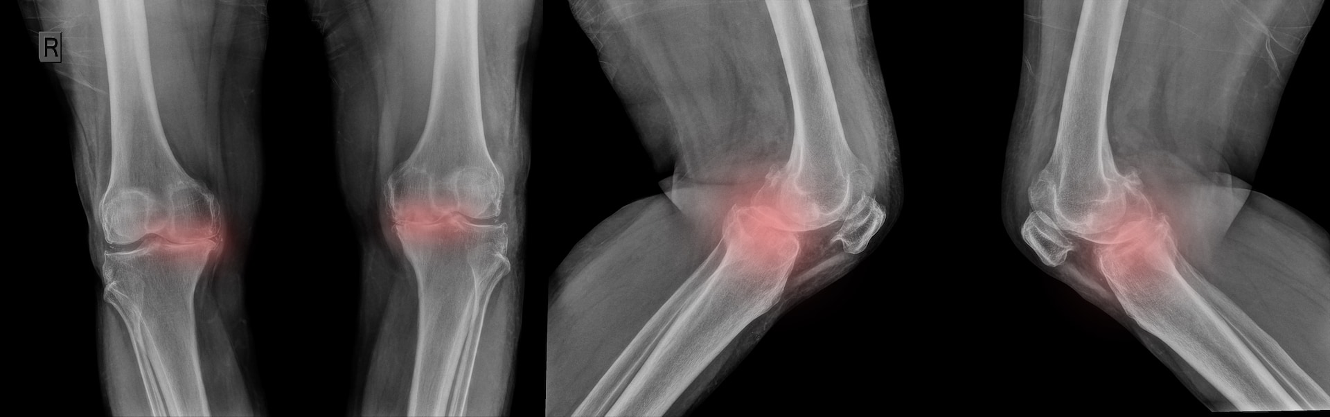
A recent paper in The Journal of Haemophilia Practice by Bhabani Sankar Dhal and colleagues describes the importance of regular assessment of joint health in people with haemophilia. Imaging technologies, like MRI scans, ultrasounds and (to a much lesser extent) X-rays, can be useful to identify changes and prevent the progression of joint disease. In this paper, the authors sadly note that in many low- and middle-income countries medical imaging equipment is in short supply.
MRI scans are the most sensitive measure and really the ‘gold standard’ for diagnosing joint problems arising from bleeding. However, the authors report that access to MRI scanners is estimated at one per million people in many Asian and sub-Saharan African countries, creating huge challenges for health care professionals looking after people with haemophilia. It is vital to be aware of any changes happening with joints so that people can have timely and appropriate treatment.
Alternative solutions
With this in mind, the authors set out to explore the possibility of creating cheaper but effective approaches to joint assessment in low-resource settings, by combining less effective imaging technologies (X-ray and ultrasound) with clinical assessment tools designed to examine joint health – such as the Hemophilia Joint Health Score (HJHS) and the Functional Independent Score in Hemophilia (FISH). These tools and scoring systems measure the health of people’s joints by looking at what a person with haemophilia can do to work out how much musculoskeletal function they have.
Over a year, the authors assessed the elbow, knee and ankle joints of participants using the HJHS and FISH scoring systems, and then used X-rays and ultrasound to generate scores for the health of the joints too. They found that combining ultrasound scoring with clinical assessment tools like the HJHS and FISH can offer a less expensive but still reliable solution to evaluate haemophilic joints.
The authors suggest that using an approach like this could enable better care for people with haemophilia in resource-constrained settings like India. However, they also recognise that if health care professionals only use these measures they may miss very early joint changes – the types of change that can only be picked up by more effective imaging techniques like MRI.
Access for all?
For me, this speaks to the theme of this year’s World Hemophilia Day: “Access for All: Prevention of bleeds as the global standard of care”. There is still a huge need to improve the accessibility of haemophilia care around the world. Finding ways to optimise care in resource constrained settings – as the researchers here have done – is obviously important work. But as a community, we also need to strive to improve access to the technologies that can be so vitally important in maintaining health for people with haemophilia, wherever they are.
Further reading
Dhal BS, Dutta A, Das A, et al. Clinical and radiological assessment of joints in people with haemophilia in Assam, Northeast India: a cross-sectional study. J Haem Pract 2023; 10(1): 11-19. doi: 10.2478/jhp-2023-0002
About the author
Rich Gorman lives with severe haemophilia A. He works as a researcher at Brighton and Sussex Medical School.
For more information about The Journal of Haemophilia Practice or Haemnet’s publishing activities, email us at publishing@haemnet.com

Scan the QR Code to follow us on Social Media
Image: Shutterstock/Somsak Thapthimthong